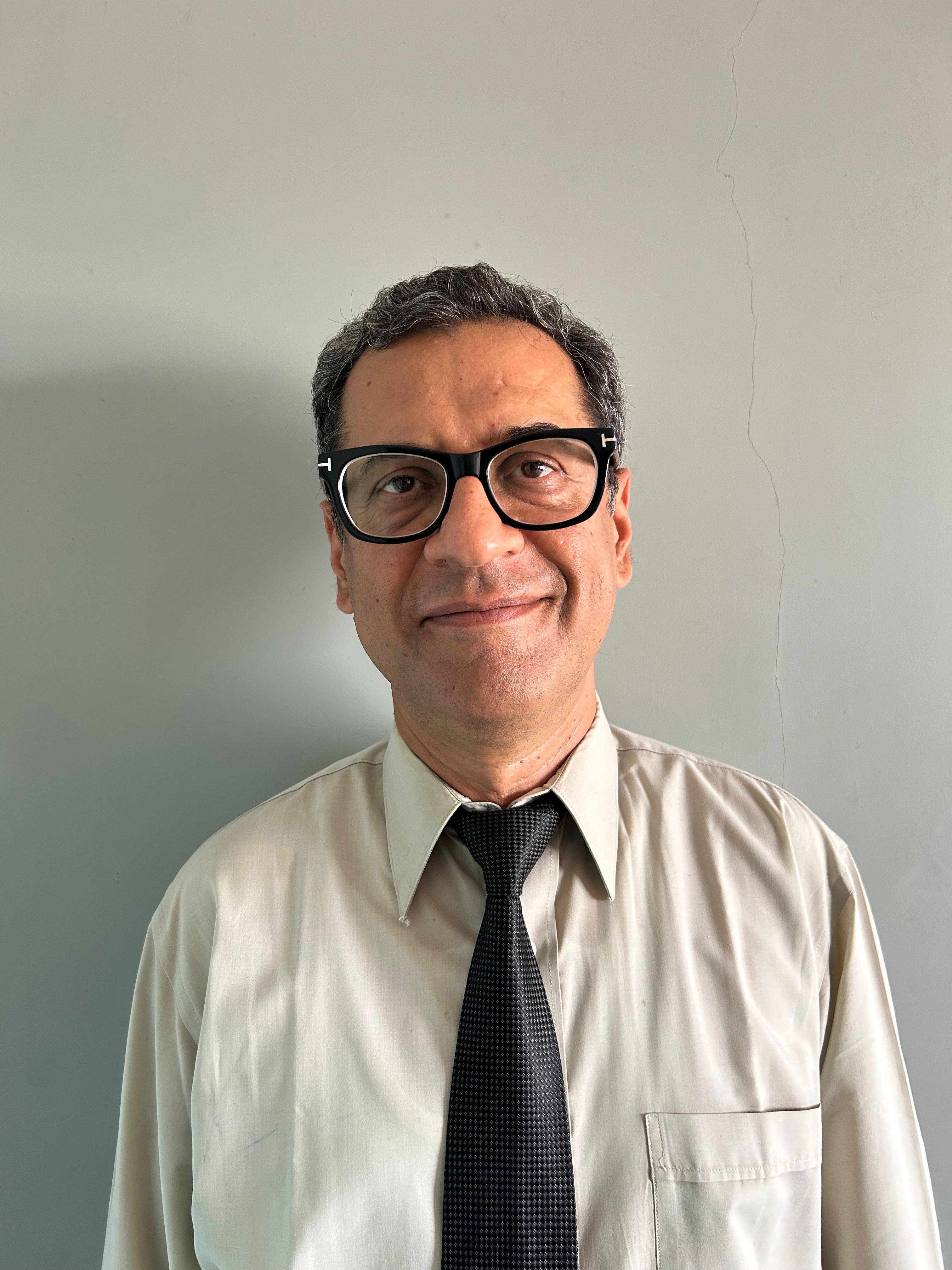
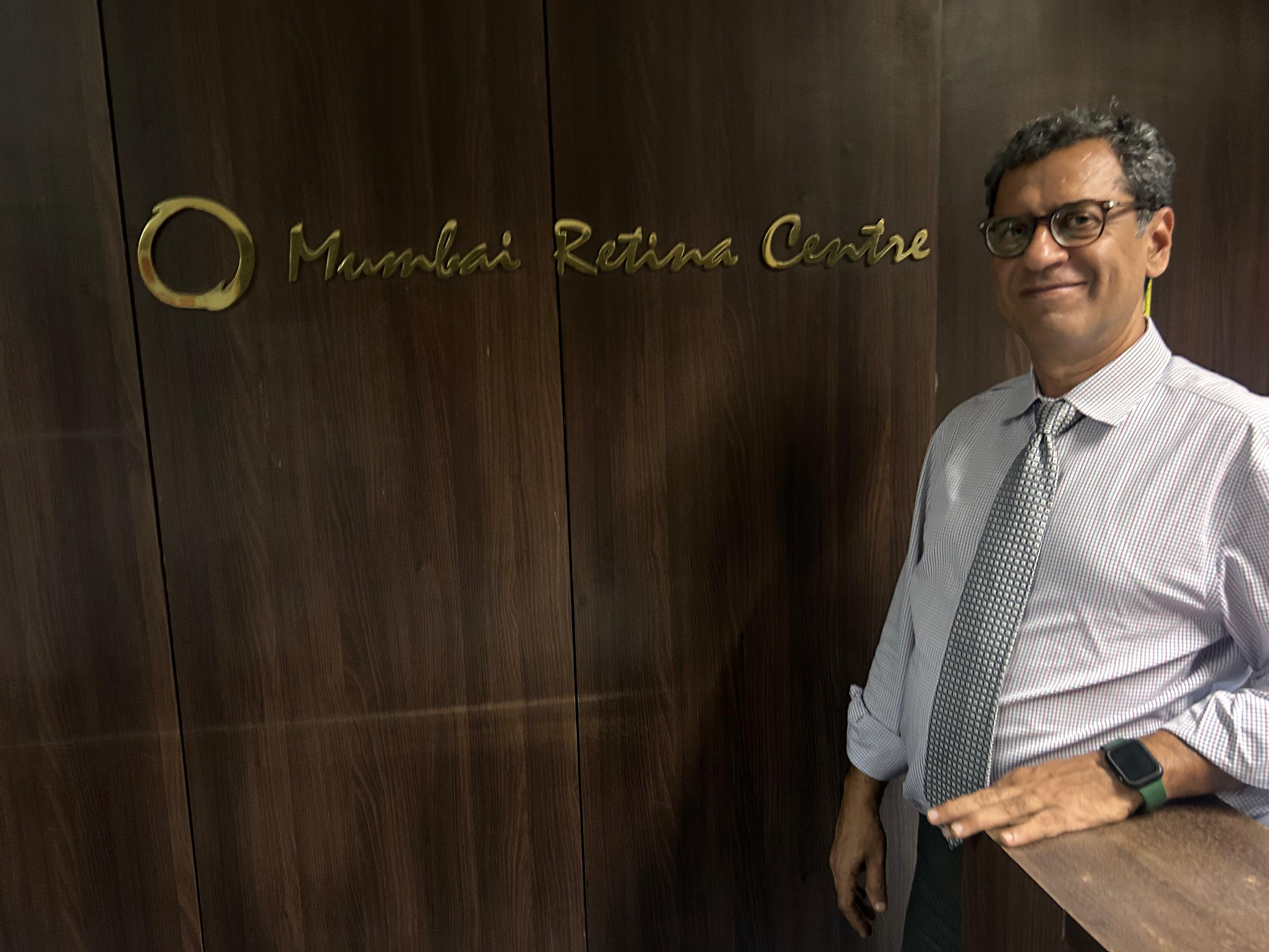
Professor Dr. Ajay I. Dudani
Vitreoretinal Specialist & Professor
Expert retinal care with international training and advanced surgical techniques
Book Consultation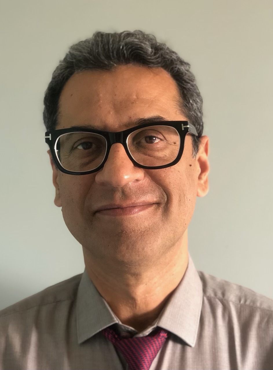
Professor Dr. Ajay I. Dudani, M.S. (Ophthalmology)
Professor • Vitreoretinal Surgeon • International Fellow
A distinguished vitreoretinal surgeon with over 30 years of experience, Professor Dr. Dudani leads Mumbai Retina Centre as a premier destination for all ophthalmic disorders and retinal diseases. His extensive international training and academic excellence position him among India's top ophthalmologists.
🎓 International Fellowships
- Sankara Nethralaya, Chennai (Dr. S.S. Badrinath)
- National Nagoya Hospital, Japan (Prof. Fumitaka Ando)
- Retina Vitreous Macula Associates, New York (Prof. Lawrence Yanuzzi)
- Doheny Eye Institute, University of Southern California
🏆 Academic Excellence
- 2nd Rank in M.S. Examination, Bombay University
- Gold Medal at D.O.M.S. Examination
- Asian Ophthalmologist Scholarship for Japan Training
- Professor of Ophthalmology, K.J. Somaiya Medical College
Virtual Tours
Explore our state-of-the-art facilities from the comfort of your home.
Mumbai Retina Centre Tour
Zen Eye Centre Operation Complex Tour
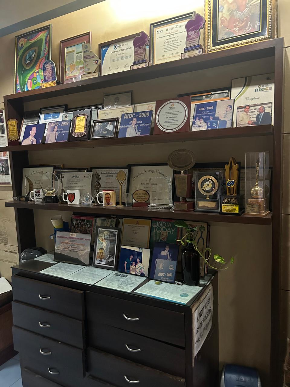
Professional Credentials & Recognition
📚 Research & Publications
Published in prestigious journals and presented at international conferences including American Academy of Ophthalmology.
🏥 Hospital Affiliations
- Professor - K.J. Somaiya Medical College & Hospital
- Sir H. N. Reliance Foundation Hospital
- Honorary Consultant - Bombay Hospital & Research Centre
- Honorary Consultant - Holy Family Hospital, Bandra
- Pramukh Swami Eye Hospital
- Navneet Jain Health Centre, Dadar
🌍 Professional Memberships
- American Academy of Ophthalmology
- All India Ophthalmology Society (AIOS)
- Vitreoretinal Society of India
- Maharashtra Ophthalmic Society
- Bombay Ophthalmic Association
🏆 Awards & Honors
- Key Opinion Leader for various ophthalmic societies and pharmaceutical companies
- Fellow of the All Indian Collegium of Ophthalmology (FACO) of the All India Ophthalmological Society (AIOS)
- On the advisory board of various pharmaceutical companies
- Mumbai Top Docs Recognition 2013, 2014 & 2015
- Asian Ophthalmologist Scholarship - Japan
- First Prize - Ophthalmology Quiz AIOS 1996
- Hargobind Medical Foundation Award
- Merit Scholarship - Bombay University
What Our Patients Say
Real stories from patients who have trusted their vision to Professor Dr. Dudani.
"Dr Dudani and the team at Mumbai Retina Eye Center are absolutely world class!!
Right from the first visit, everybody was helpful. I truly felt that Dr Ajay was completely focused on giving me the best care, his experience really shows..
I visited for a consultation on Lasik treatment, and Doctor was very caring and took the time to explain the entire process step by step. This is very important since I was quite scared of getting a procedure in my eye. Hats off to the entire team, I will only come here for my eye care!! No other clinic in Mumbai (or even in India) can compare."
"Dudani sir is somehow the absolute best at everything he does. He's not only an exceptional ophthalmologist but also someone I blindly trust. His level of compassion and genuine care for patient truly sets him apart. His kindness and dedication go far beyond the clinic. Highly recommend!!"
"Had recently visited Dr. Ajay Dudani at Mumbai Retina Center for Keratoconus. His expertise in surgery is second to none and his cheerful and friendly behaviour towards patients is one of his best traits.
Have already recovered fully and am feeling great.
Thanks to the team at Mumbai Retina Centre!❤️"
Our Services
General Conditions
A cataract is a clouding of the normally clear lens of the eye, often compared to a frosted or yellowed window. Common symptoms include fuzzy vision, glare, poor night vision, and fading colors. While most common with aging, it can also be caused by diabetes, injury, or medications. Treatment is a highly successful surgery to replace the cloudy lens.
Learn MoreChoosing the right Intraocular Lens (IOL) is key for cataract surgery. The best choice depends on your lifestyle and vision needs.
- Monofocal IOLs: Offer clear vision at a single distance (near, intermediate, or far). Best for patients comfortable with glasses for other distances.
- Multifocal IOLs: Provide clear vision at multiple distances, reducing the need for glasses. May cause halos or glare at night.
- Toric IOLs: Correct astigmatism, providing clearer vision for those with an irregular corneal surface.
- EDOF IOLs: Offer a balanced range of vision with fewer side effects than multifocal lenses, ideal for active lifestyles.
A consultation is crucial to determine the most suitable lens for you.
Learn MoreLASIK (Laser-Assisted in Situ Keratomileusis) is a popular refractive surgery that corrects myopia (nearsightedness) to reduce or eliminate the need for glasses and contact lenses. We utilize advanced Wavefront Guided Custom LASIK, which analyzes your eye's unique properties to create a personalized treatment. This approach offers a higher potential for 20/20 vision and reduces side effects like night glare.
Learn MoreGlaucoma is a serious condition that damages the optic nerve, often due to increased pressure in the eye. It is a leading cause of irreversible blindness, but early detection and treatment can prevent vision loss. Since it often has no early symptoms, regular eye exams, especially after age 40, are critical. Treatment involves managing eye pressure through medication, laser, or surgery.
Learn MoreUveitis is the inflammation of the uvea, the eye's middle layer. It can cause significant damage and vision loss if left untreated. Symptoms include eye redness, pain, light sensitivity, and blurred vision. Uveitis can be linked to infections, injuries, or systemic diseases, but often the cause is unknown. A "red eye" should never be ignored and requires prompt evaluation by an ophthalmologist.
Learn MoreDry eye occurs when your eyes don't produce enough quality tears. Symptoms include stinging, burning, scratchiness, and even excessive tearing. It's often linked to aging, hormonal changes, or medications.
Computer Vision Syndrome (CVS) is a major contributor today, caused by prolonged screen use. We provide diagnosis and management for dry eye, including tips to reduce CVS symptoms like the 20-20-20 rule and specialized eye drops.
Learn MoreThis specialty focuses on the eyelids, tear ducts, and the orbit (the bony eye socket). We treat conditions like ptosis (drooping eyelids), eyelid malpositions, blocked tear ducts, orbital fractures, and tumors. This also includes cosmetic procedures to enhance the appearance of the eyes and surrounding areas.
Learn MoreNeuro-ophthalmology bridges neurology and ophthalmology, addressing vision problems related to the nervous system. This includes disorders of the optic nerve, visual pathways, and brain, such as optic neuritis, double vision, and vision loss from strokes or tumors. A comprehensive evaluation is crucial for diagnosing these complex conditions.
Learn MoreRetinal Diseases
A common complication of diabetes, this condition affects blood vessels in the retina. It is a leading cause of blindness in working-age adults. Early detection and treatment are critical.
Key Treatments:
Learn MoreA medical emergency where the retina pulls away from its normal position. Symptoms include sudden floaters, flashes of light, or a curtain-like shadow over your vision. Immediate treatment is required to save vision.
Key Treatments:
Learn MoreAge-related macular degeneration (AMD) is a leading cause of vision loss in older adults. It affects central vision, making it difficult to read, drive, or recognize faces. We treat both "dry" and "wet" forms of AMD.
Key Treatments:
Learn MoreA small break in the macula, the part of the retina responsible for sharp, detailed central vision. This can cause blurred and distorted vision. Macular holes are often repaired with vitreoretinal surgery.
Key Treatments:
Learn MoreThis occurs when a vein in the retina is blocked, leading to blood and fluid leakage into the retina. It can cause sudden vision loss and requires prompt treatment to prevent complications like macular edema and neovascular glaucoma.
Key Treatments:
Learn MoreWhile often harmless, a sudden onset of floaters and flashes can signal a serious condition like a retinal tear or detachment. A thorough examination is necessary to rule out any underlying problems and determine the appropriate course of action.
Learn MoreMeet Our Associate Doctor

Dr. Anadya Dudani
Ophthalmologist • MBBS (Distinction)
Dr. Anadya Dudani is a budding ophthalmic surgeon presently working at Rajawadi Hospital Mumbai. She has passed her MBBS from KJ Somaiya Medical College and Hospital with distinction in 9 subjects with flying colours.
Ever since her school days, she pictured herself as an ophthalmologist just like her father, and now she works everyday to make that dream come true under a microscope, performing cataract surgeries.
Educational Background
- MBBS: KJ Somaiya Medical College and Hospital (Distinction in 9 subjects)
- Schooling: ICSE from Dhirubhai Ambani International School and Arya Vidya Mandir Bandra
Sports & Interests
Dr. Anadya is an avid sportsperson with interests in pickleball, tennis, swimming, and deep sea diving.
Current Practice
Position: Ophthalmologist at Rajawadi Hospital Mumbai
Specialization: Cataract Surgery and General Ophthalmology
Mumbai Ultrasound Centre

Dr. Anupam A. Dudani
Senior Radiologist • 25+ Years of Experience
Dr. Anupam A. Dudani, a senior radiologist with 25 years of experience, is the head and founder of Mumbai Ultrasound Centre. She has been practicing at Hinduja Hospital, Khar since its inception.
Areas of Specialization
- Obstetrics Sonography, Anomaly Scan
- Mammography and Elastography
- Female Imaging and Neonatal Sonography
Expertise
Dr. Dudani is adept at Color Doppler and ultrasound-guided biopsies.
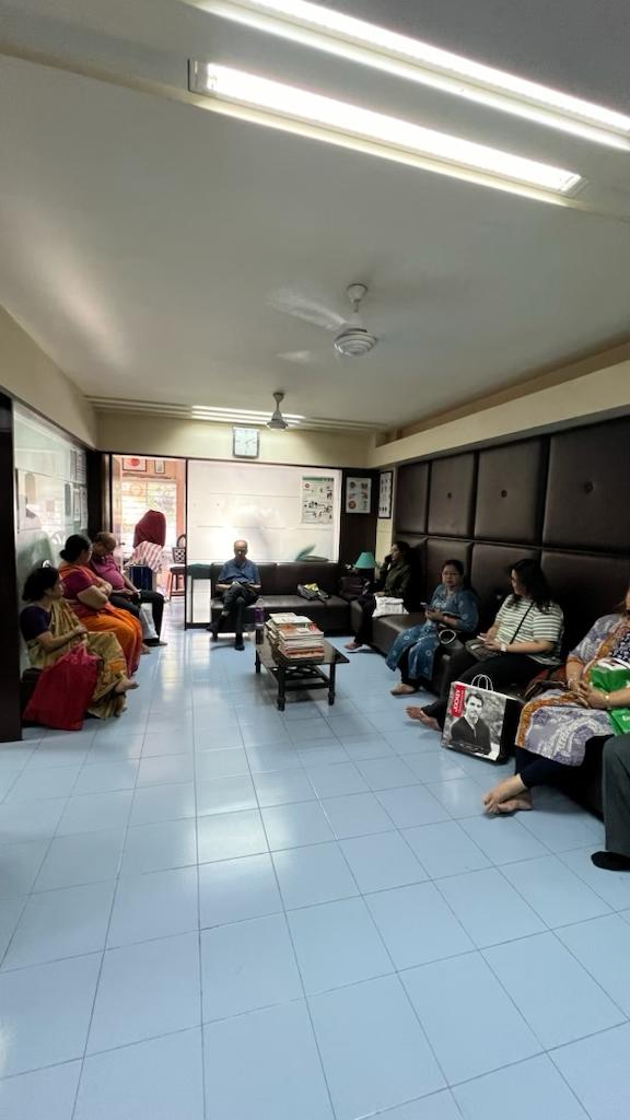
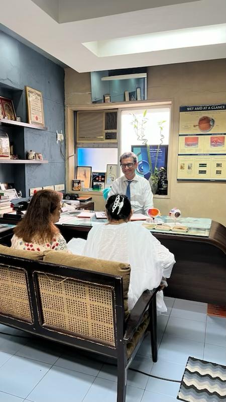
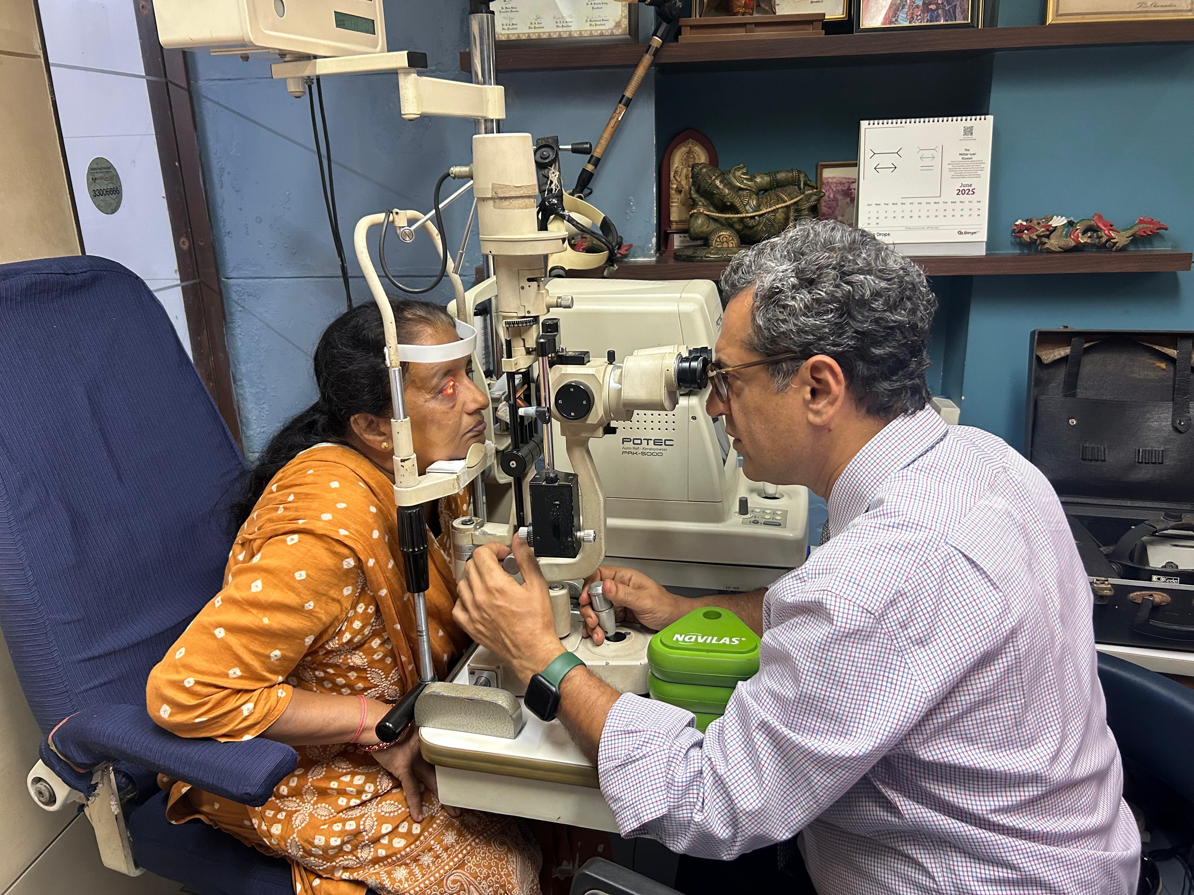
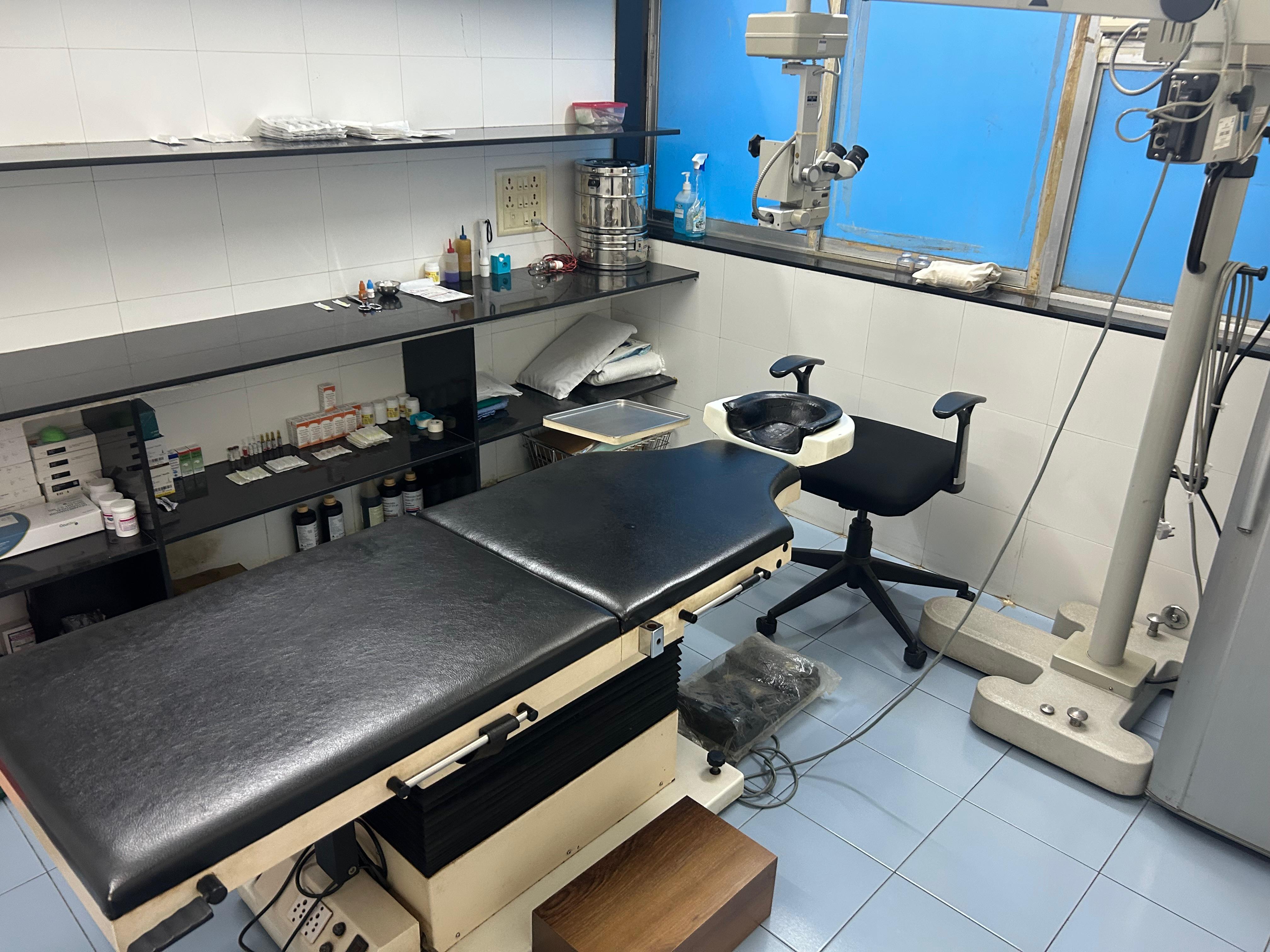
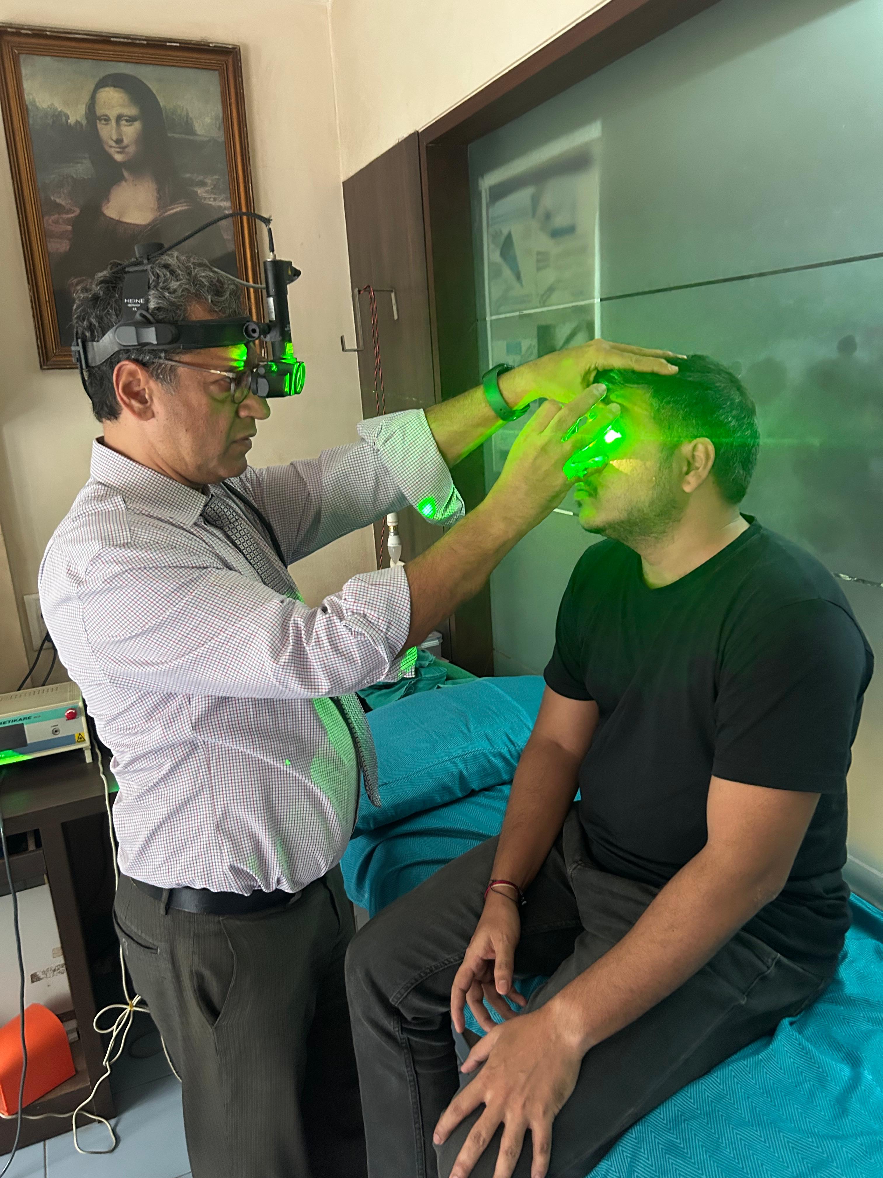
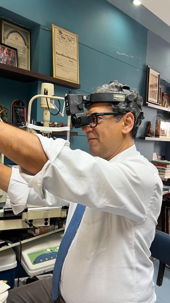
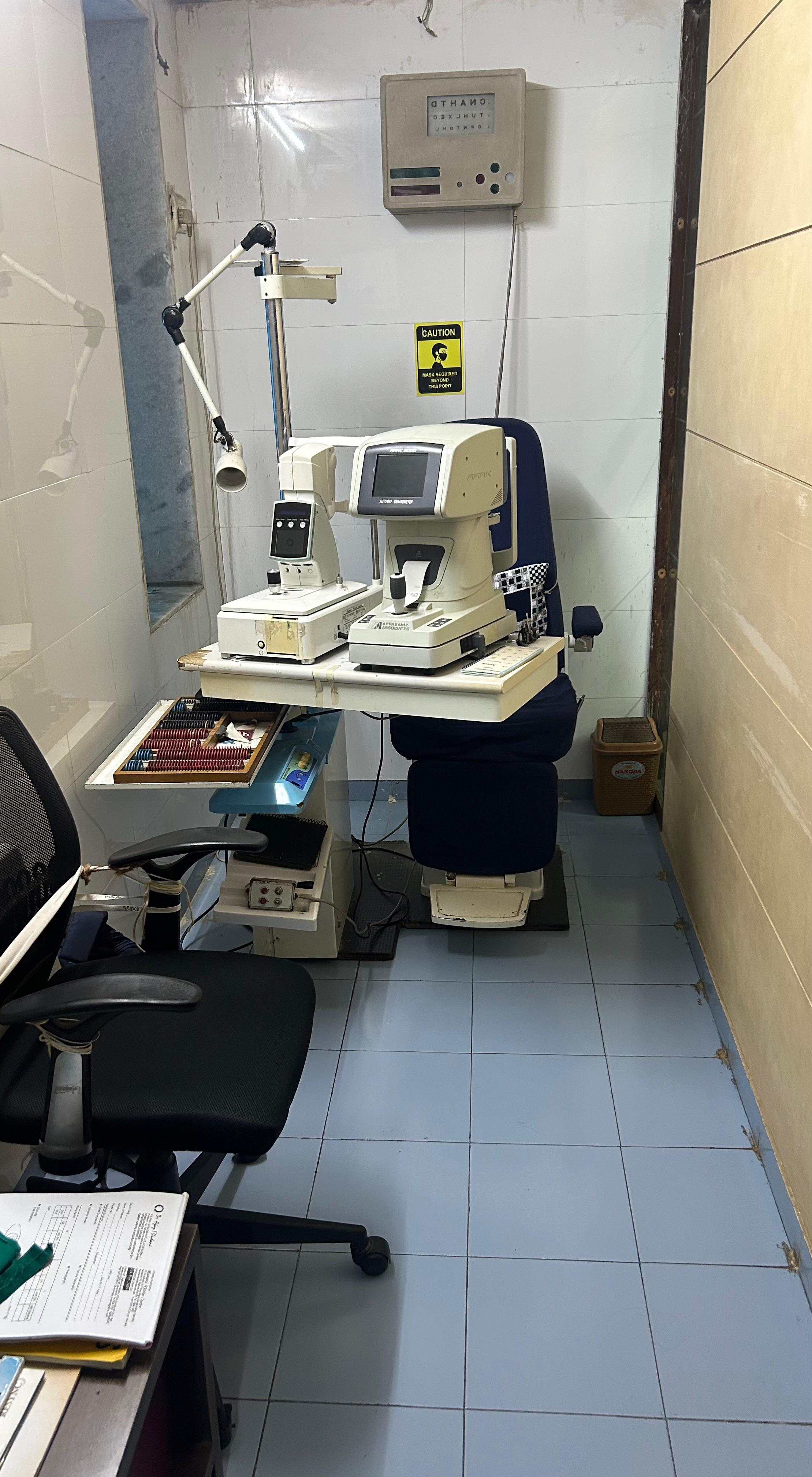
Clinic Gallery
State-of-the-art facilities and advanced medical equipment
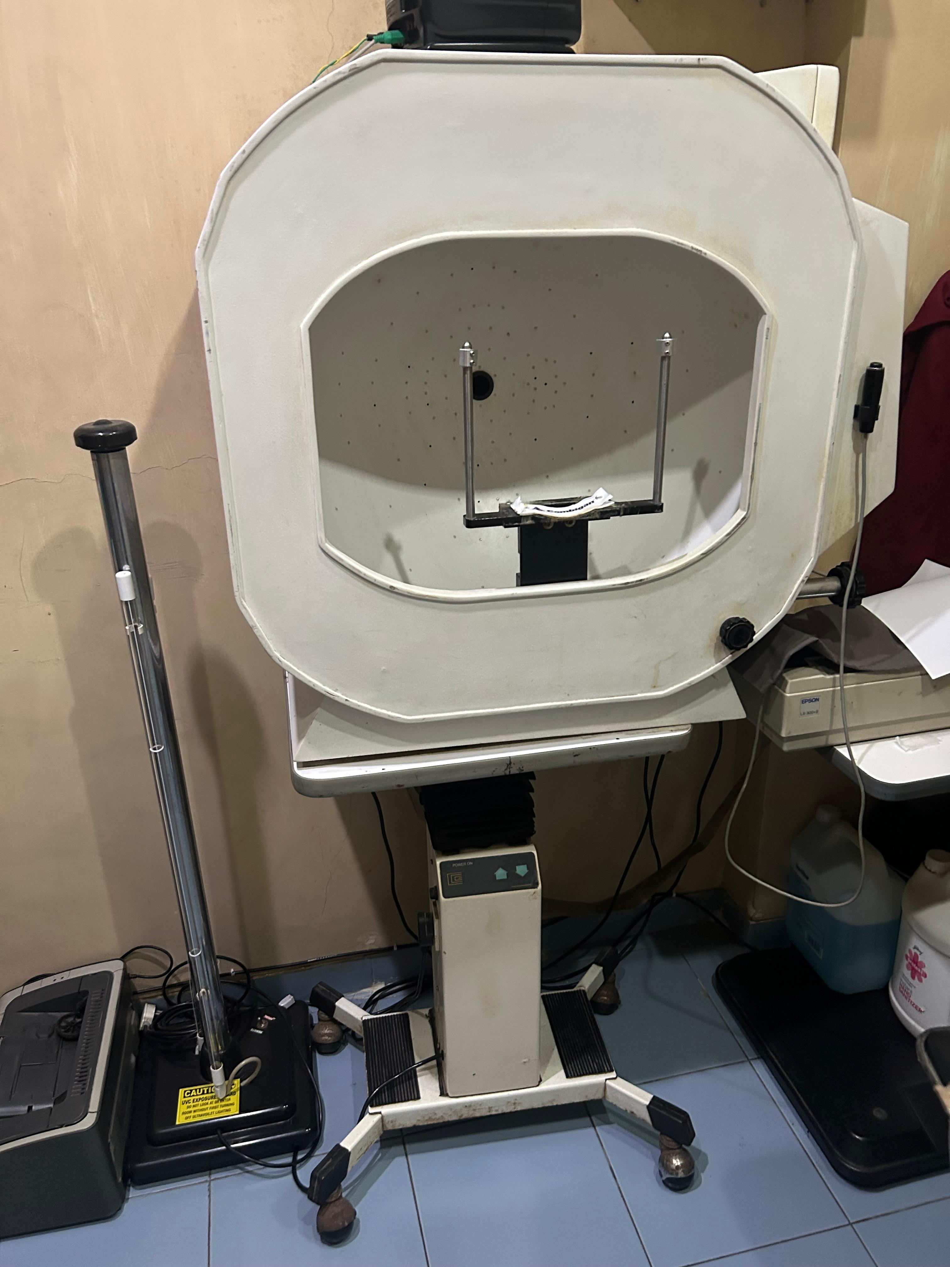
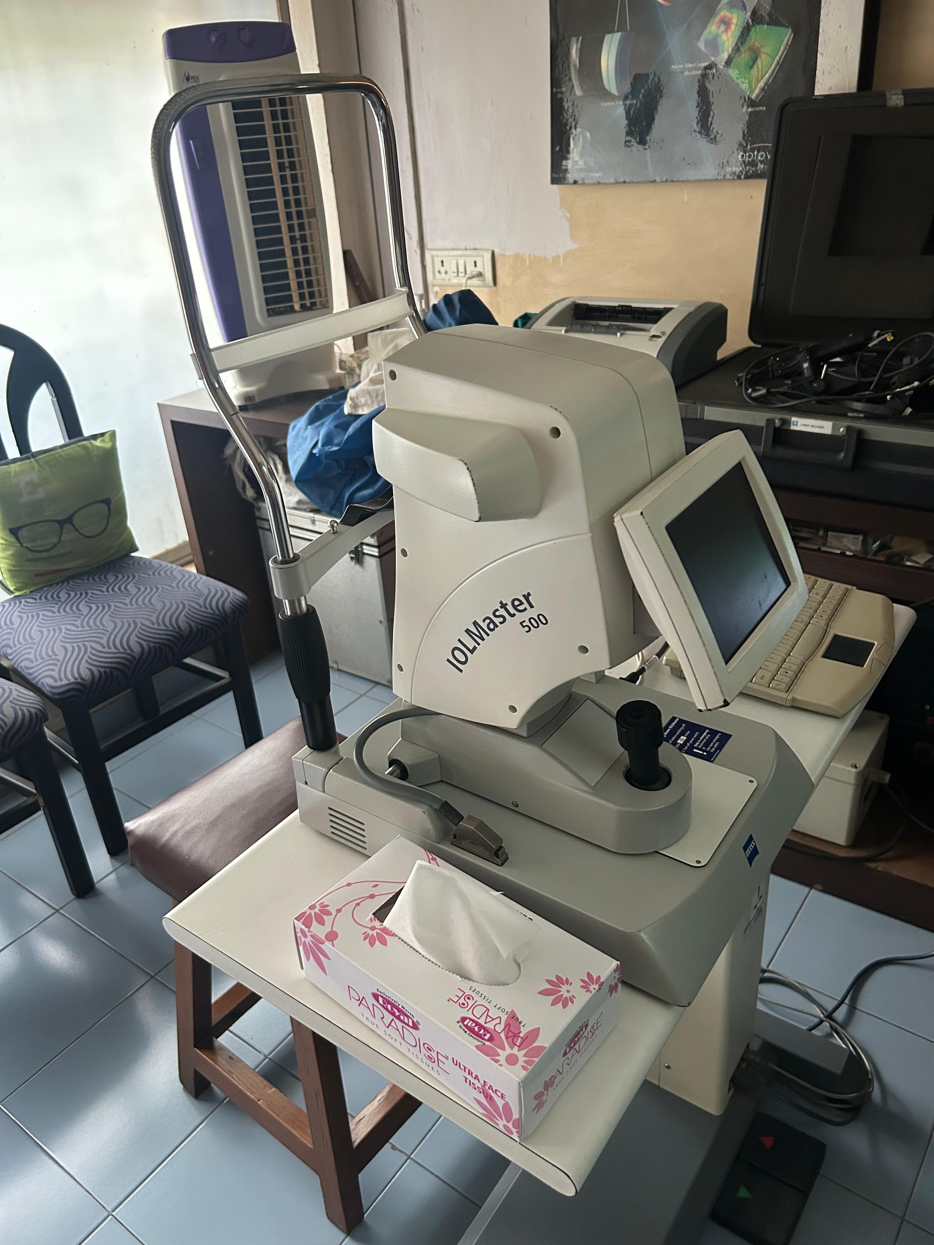
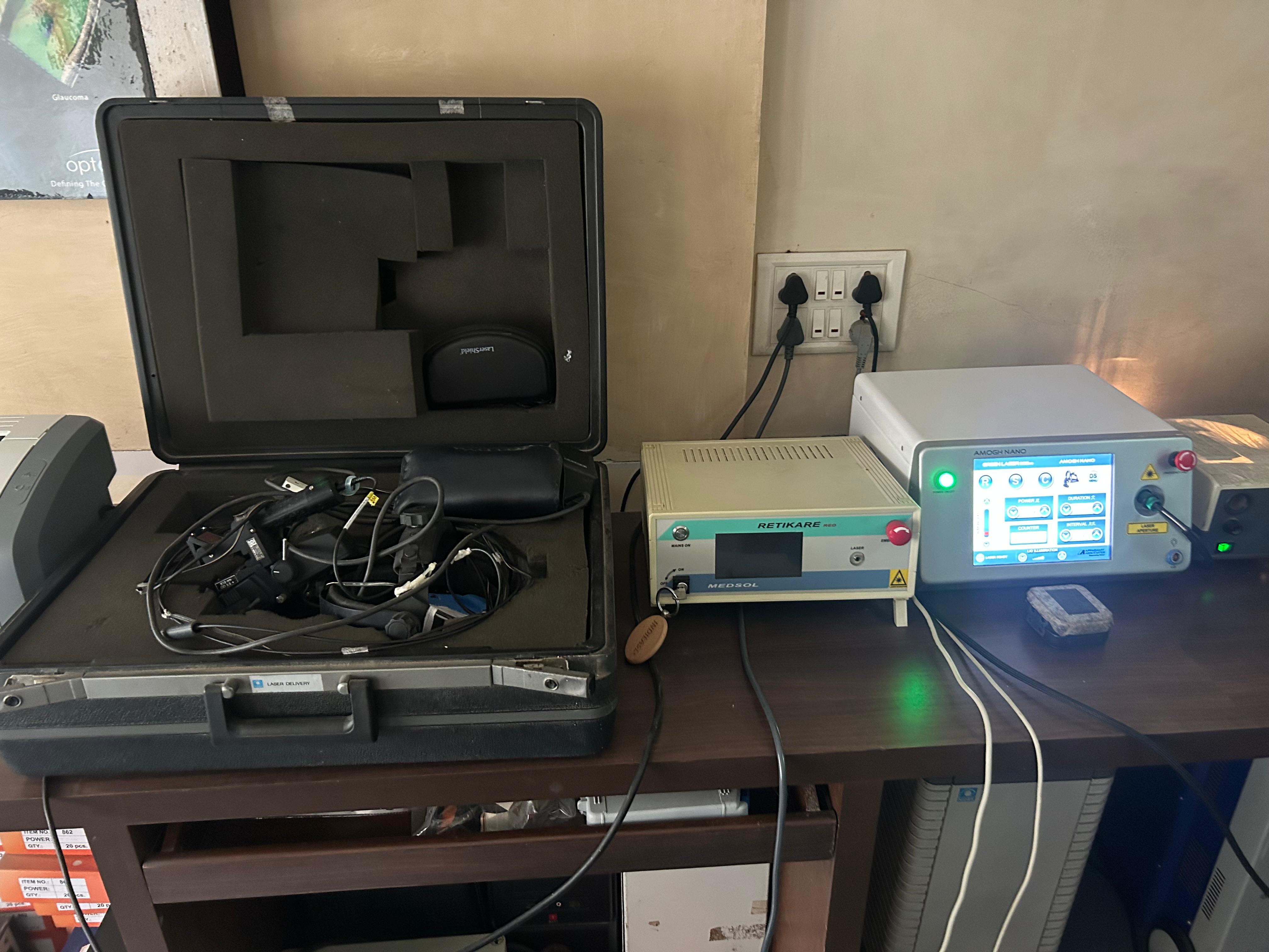
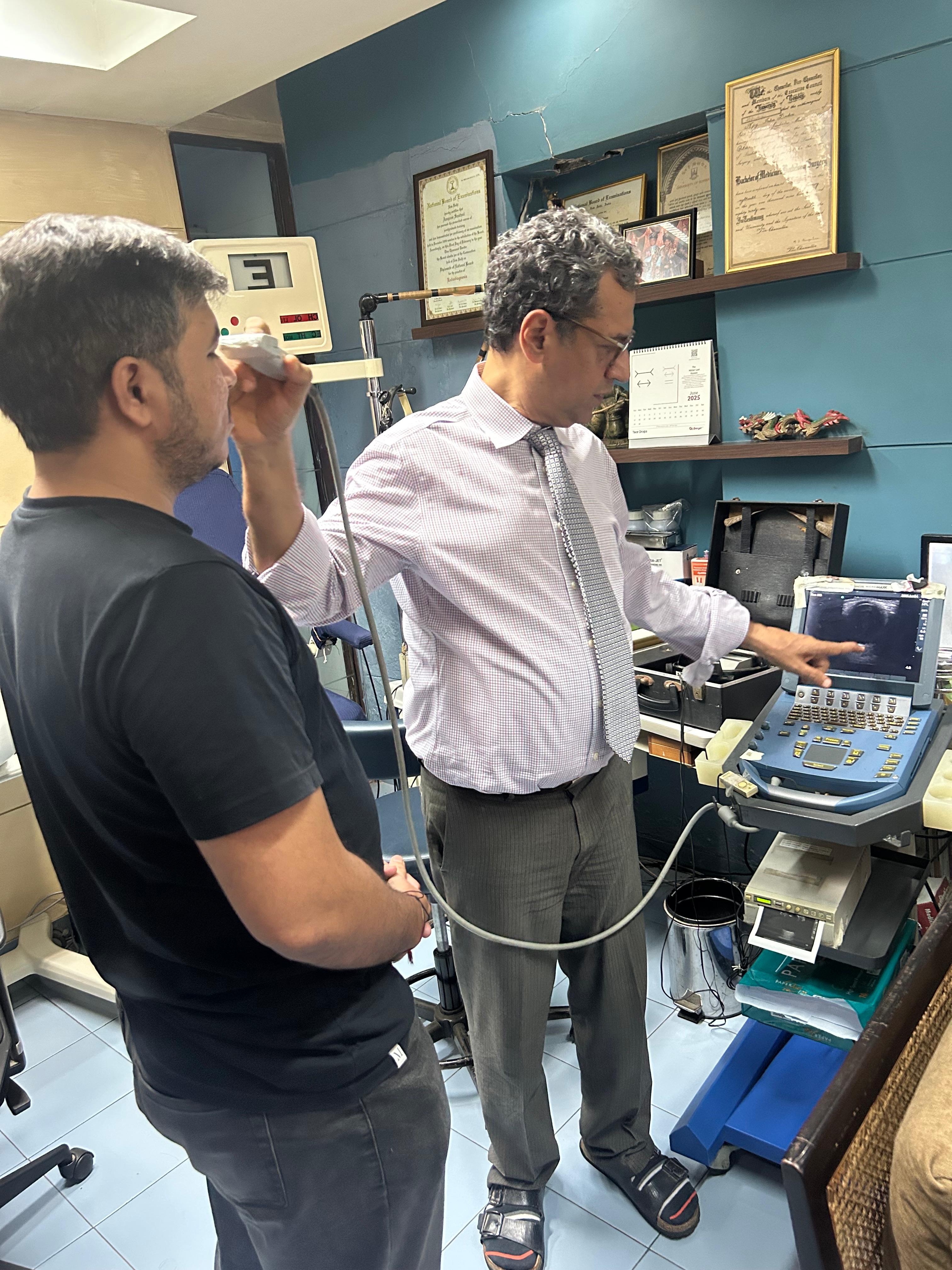
Clinic Facilities
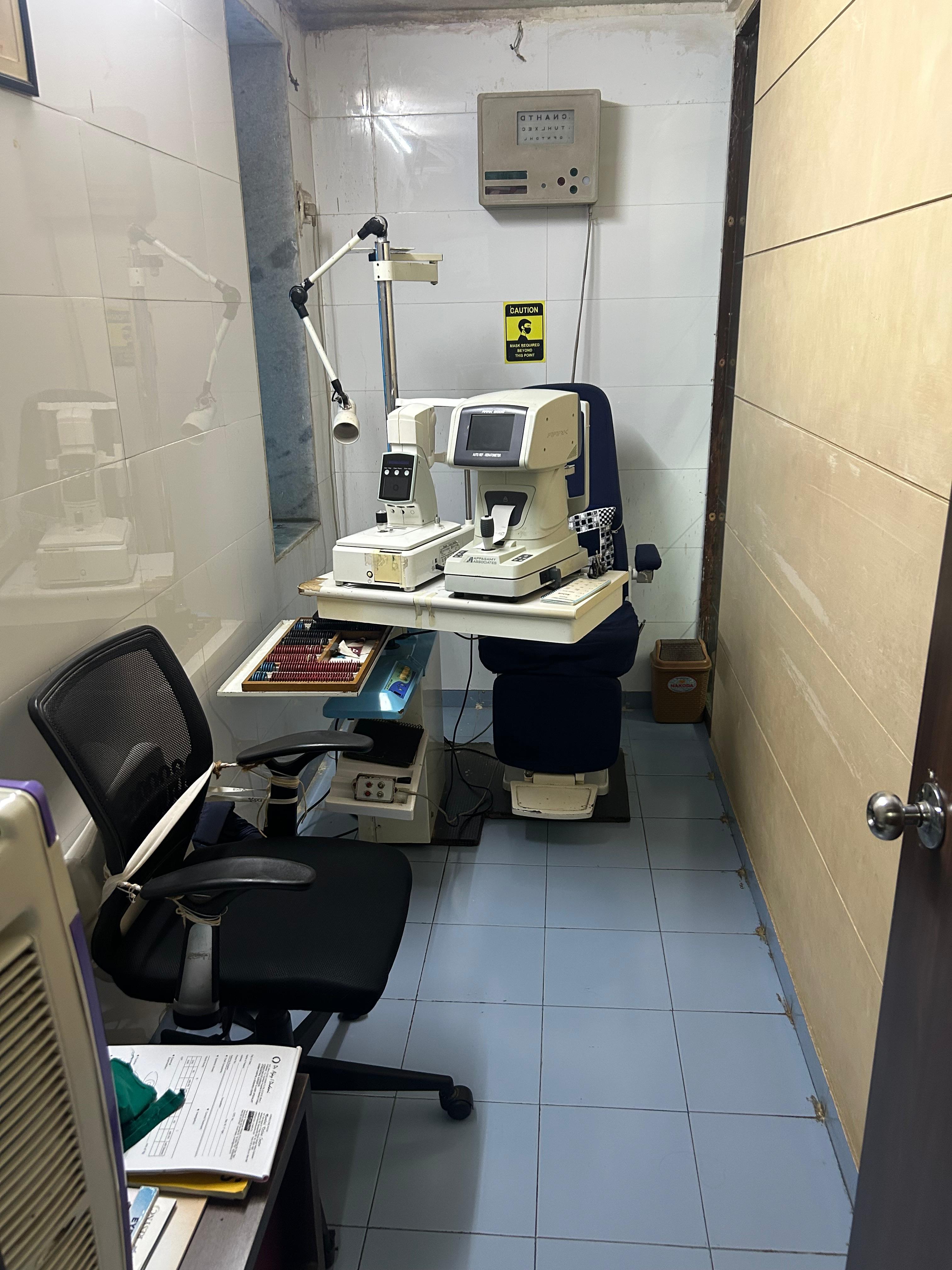
Procedures & Equipment
.jpg)
Frequently Asked Questions
Expert answers to common questions about retinal conditions, treatments, and surgical procedures.
Retinal detachment is caused by a combination of factors including retinal holes, retinal breaks or retinal tears, liquefaction of the vitreous humor, and mechanical forces on the retina (traction). The most common causes include:
- Age-related changes: Natural aging process causing vitreous shrinkage
- High myopia: Elongated eyeball stretching the retina
- Previous eye surgery: Cataract surgery can occasionally lead to complications
- Eye trauma: Blunt force or penetrating injuries
- Diabetic retinopathy: Advanced diabetes causing scar tissue formation
Retinal detachment can lead to severe vision loss or blindness if left untreated, but with prompt surgical intervention, many patients maintain good vision. The outcome depends on:
- Location: Detachments involving the macula (central vision) have more impact
- Duration: Earlier treatment leads to better outcomes
- Extent: Partial vs. complete detachment
- Cause: Some types respond better to treatment than others
Emergency surgical repair can often restore and preserve vision when performed promptly.
High myopia (severe near-sightedness) significantly increases the risk of retinal detachment. This occurs because:
- The elongated eyeball stretches the retina, making it thinner
- Peripheral retinal degeneration is more common
- Vitreous changes occur earlier in myopic eyes
- The risk increases with the degree of myopia
Recommendation: Patients with high myopia should have regular retinal examinations to detect early warning signs.
Modern retinal detachment repair utilizes several advanced surgical techniques:
Vitrectomy
Removal of vitreous gel and repair of retinal tears using gas or oil tamponade
Scleral Buckle
External support placed around the eye to close retinal breaks
Pneumatic Retinopexy
Gas bubble injection combined with laser or freezing treatment
Laser/Cryotherapy
Sealing retinal tears to prevent further detachment
Diabetic patients require regular retinal screening based on their diabetes type and duration:
- Type 1 Diabetes: Annual exams starting 5 years after diagnosis
- Type 2 Diabetes: Annual exams starting at diagnosis
- Pregnancy with Diabetes: Exams each trimester
- Advanced Retinopathy: Every 3-6 months or as recommended
Early detection and treatment can prevent 90% of severe vision loss from diabetes.
Diabetic retinopathy progresses through distinct stages:
Mild Non-Proliferative
Small areas of balloon-like swelling in retinal blood vessels (microaneurysms)
Moderate Non-Proliferative
Blood vessels begin to swell and lose ability to transport blood
Severe Non-Proliferative
More blood vessels blocked, areas of retina lose blood supply
Proliferative Diabetic Retinopathy
Growth of new abnormal blood vessels and scar tissue
While diabetic retinopathy cannot be completely prevented, its progression can be significantly slowed or stopped through:
- Blood Sugar Control: Maintaining HbA1c below 7%
- Blood Pressure Management: Target below 140/90 mmHg
- Cholesterol Control: Managing lipid levels
- Regular Exercise: Improving overall cardiovascular health
- Smoking Cessation: Eliminating additional vascular risk
- Regular Eye Exams: Early detection and treatment
Modern vitreoretinal surgery has excellent success rates:
- Retinal Detachment: 85-95% anatomical success rate in primary surgery
- Macular Hole: 90-95% closure rate with vision improvement
- Diabetic Vitrectomy: 70-85% stabilization or improvement
- Epiretinal Membrane: 85-90% visual improvement
Success depends on factors like timing of surgery, extent of damage, and patient's overall eye health.
Surgery duration varies based on complexity:
- Simple Retinal Detachment: 1-2 hours
- Complex Vitrectomy: 2-4 hours
- Macular Hole Repair: 1-1.5 hours
- Diabetic Vitrectomy: 2-3 hours
Most procedures are performed on an outpatient basis under local or general anesthesia.
Recovery varies by procedure but generally includes:
First 24-48 Hours
- Eye patch and shield protection
- Limited activity and rest
- Prescribed eye drops
- Possible positioning requirements
First Week
- Gradual return to light activities
- Continued eye drop regimen
- Avoid heavy lifting or straining
- Follow-up appointments
2-6 Weeks
- Progressive activity increase
- Vision gradually improves
- Continue protective measures
- Regular monitoring visits
Seek immediate medical attention if you experience:
🚨 Immediate Emergency
- Sudden, severe vision loss
- Curtain or shadow across vision
- Sudden onset of many new floaters
- Flashing lights with vision changes
⚠️ Schedule Soon
- Gradual vision changes
- New onset of floaters
- Distorted central vision
- Difficulty with night vision
While retinal surgery is generally safe, potential complications include:
- Infection: Less than 1% risk with proper sterile technique
- Cataract Formation: May accelerate existing cataracts
- Increased Eye Pressure: Temporary or rarely permanent
- Re-detachment: 5-15% may require additional surgery
- Double Vision: Usually temporary from positioning
Dr. Dudani's extensive experience and advanced techniques minimize these risks significantly.
While a referral from your primary eye doctor is helpful, it's not always required:
- Emergency Situations: No referral needed for urgent symptoms
- Insurance Requirements: Some plans may require referrals
- Second Opinions: Always welcome and encouraged
- Routine Screening: High-risk patients can self-refer
Contact our office to discuss your specific situation and insurance requirements.
Schedule Your Consultation
Send us a Message
Have a question or want to schedule a visit? Fill out the form below.